Nephrology
Pulmonology
Robot-Assisted Surgery
Urology
Book an appointment
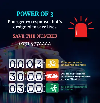
0731-4774444
Emergency
0731 4774129
Follow Us
Consult Super-Specialist Doctors at CARE Hospitals
Health Packages
Book an Appointment
Call Us
Get A Call Back From Our Health Advisor Now
Enter your details, and our advisor will call you back shortly!