A PET-CT scan is an investigation performed when a doctor needs to understand how metabolically active a tissue is, whether a tumour is responding to treatment, or whether cancer has spread beyond the primary site. It combines two imaging technologies in a single examination, giving oncologists and doctors a level of diagnostic detail that neither modality alone can match.
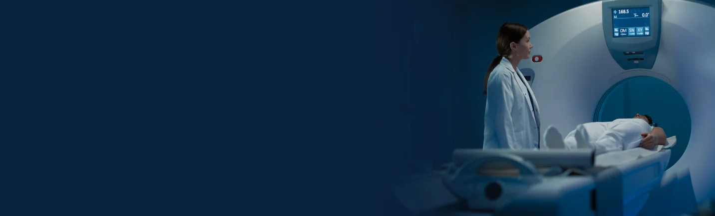
At Ramkrishna CARE Hospitals, Raipur, PET-CT scanning is available through the United Imaging uMI 550 – a digital PET-CT system combining a 24 cm axial FOV PET detector with an 80-slice CT, delivering high resolution and high sensitivity. One of the most advanced PET CT platforms in Central India, available to patients across Chhattisgarh.
What is a PET-CT scan?
PET CT combines two imaging technologies in a single session. PET (Positron Emission Tomography) captures cellular metabolism, and CT (Computed Tomography) provides detailed anatomical maps. The fused image shows both simultaneously: where structures are and what they are doing metabolically.
The PET component uses FDG (a glucose analogue). Cancer cells, being metabolically active, take up more tracer and appear as areas of increased signal. The CT component maps the precise anatomical location of those signals.
The uMI 550 uses an integrated light guide digital PET detector with SiPM technology, achieving time-of-flight imaging with 2.76 mm crystal pixel resolution. CT images are reconstructed at up to a 1024×1024 matrix, revealing small lymph nodes, early lesions and subtle changes that lower-resolution systems miss.
Why is a PET-CT scan performed?
A PET CT is ordered when a doctor needs information about how tissues are functioning, not just what they look like. Common scenarios are the following:
- Cancer staging: It helps determine disease extent, including lymph node and distant organ involvement, before treatment planning.
- Treatment response assessment: Evaluating whether chemotherapy, radiation or targeted therapy is achieving the intended effect
- Cancer recurrence detection: Identifying the return of disease even before it is visible on CT or MRI
- Radiation therapy planning: Delineating the metabolically active tumour volume for radiotherapy target definition.
- Neurological investigation: Brain function evaluation in epilepsy, dementia, and movement disorders.
- Cardiac viability: Determining whether reduced heart muscle retains viable tissue suitable for revascularisation.
- Infection and inflammation: Identifying the source of fever of unknown origin.
Conditions Diagnosed with PET CT Scan
A PET scan in Raipur is used to diagnose various conditions. They are:
- Oncological Conditions: These include:
- Lung cancer: Staging and recurrence surveillance
- Lymphoma (Hodgkin and non-Hodgkin): Staging, treatment response and end-of-treatment evaluation
- Colorectal cancer: Preoperative staging and recurrence detection
- Head and neck cancers: Tumour localisation and lymph node staging
- Breast cancer: Staging in selected high-risk presentations and recurrence evaluation
- Oesophageal and gastric cancer: Staging before surgery or definitive chemoradiation
- Melanoma: Staging and detection of metastatic disease
- Thyroid cancer: Recurrence detection in patients with elevated thyroglobulin & negative conventional imaging
- Cervical and ovarian cancers: Staging and restaging
- Brain tumours: Differentiation of recurrence from radiation necrosis
- Unknown primary tumour: Identification of the primary site when metastatic disease has been identified without a clear source.
- Non-oncological conditions
- Epilepsy: Identification of the seizure focus for surgical planning
- Neurodegenerative disorders: Alzheimer's disease and Parkinson's disease evaluations.
- Cardiac viability: Myocardial viability assessment in ischaemic heart disease
- Fever of unknown origin: Localisation of an occult infection or inflammatory focus.
Types of PET CT Scans
The type of PET CT scan performed depends on the clinical question being answered. The most common variants include:
- Whole-body FDG PET-CT: The standard oncological staging scan, covering from the skull base to mid-thigh or the full body (using fluorodeoxyglucose as the tracer)
- Limited-field FDG PET CT: Focused imaging of a specific anatomical region where whole-body coverage is not required
- Brain FDG PET CT: Dedicated high-resolution imaging of brain metabolism for neurological evaluation
- Cardiac PET-CT: Myocardial perfusion & viability assessment, typically using different tracers
- 68Ga PSMA PET CT: Used in prostate cancer staging and recurrence detection, with the PSMA ligand targeting prostate-specific membrane antigen
- 68Ga DOTATATE PET CT: Used for neuroendocrine tumour staging & therapy planning
The uMI 550 supports all protocols. Its 240 mm axial FOV and 11 cps/kBq system sensitivity complete whole-body scans in four bed positions and are faster than conventional six-bed systems, maintaining full diagnostic quality.
PET CT Scan Procedure
Understanding what happens during the scan helps patients approach the examination with confidence rather than anxiety. The procedure follows a structured sequence:
- Arrival and registration: Clinical history taken, contraindications confirmed
- Tracer injection: FDG administered through a small IV cannula. The radioactivity dose is very small and well within established safety parameters.
- Uptake period: 60 minutes of quiet rest while the tracer distributes itself. Patients are asked to remain still and avoid talking to minimise uptake in non-target tissues.
- Scanning: Approximately 15 to 30 minutes on the PET-CT table. The uMI 550's EasyScan system automatically adjusts the CT range and PET bed positions for each patient's anatomy.
- Completion: Patients leave immediately after scanning. Normal diet and activity resume straight away.
Preparation Before PET CT
Preparation steps are the following:
- Fast for 4 to 6 hours before the scan, as food intake raises blood glucose levels, which competes with FDG uptake in tumour tissue & reduces image quality. Water is permitted and encouraged.
- Avoid vigorous physical exercise for 24 hours before the scan – exercise increases glucose uptake in muscles, which can interfere with image interpretation.
- Inform the team of any diabetes medication, as diabetic patients require specific preparation protocols to optimise blood glucose levels before injection.
- Avoid caffeine and alcohol for 24 hours before the scan.
- Inform the team of any recent surgery, radiotherapy or chemotherapy, as these can affect image interpretation.
- Arrive wearing comfortable, loose clothing without metal zips or fasteners.
- Bring all previous imaging reports, scans, and clinical letters to the appointment
- Inform the team if pregnant or breastfeeding, as PET-CT uses ionising radiation and a radiotracer, and specific guidance applies in these situations.
Benefits of a PET-CT Scan
Key advantages are:
- Whole-body staging in a single examination: Cancer staging that previously required multiple separate investigations can be completed in one PET-CT session.
- Detection of metabolically active disease before anatomical changes are visible: PET can identify areas of malignant activity when the lesion is still too small to create a visible mass on a CT or MRI.
- Accurate treatment response assessment: Changes in metabolic activity after treatment often precede measurable changes in tumour size, allowing earlier determination of whether a treatment is working.
- High sensitivity for recurrence detection: PET-CT detects cancer recurrence with greater sensitivity than CT alone, particularly in sites where post-treatment fibrosis or scarring can obscure conventional imaging.
- Radiation therapy target optimisation: PET CT-defined tumour volumes more accurately reflect the extent of metabolically active disease, allowing radiation dose to be concentrated where it is most needed.
- Lower radiation dose compared to multiple separate scans: A single PET-CT examination replaces what might otherwise require multiple investigations with a combined radiation burden.
Why Choose Ramkrishna CARE Hospitals for a PET-CT Scan in Raipur?
PET CT quality is determined by three things: the technology, the expertise interpreting results, and the clinical context. Ramkrishna CARE Hospitals provides all three.
- United Imaging uMI 550 Digital PET CT: ILG Digital PET detector with SiPM technology, 80-slice CT with Z-Detector, 0.55 mm pixel acquisition, 2.9 mm NEMA resolution with Time-of-Flight, and lesion detection as small as 3.5 mm at 0.085 mCi/kg.
- Digital Self-Gating: Automatically detects respiratory motion without external devices, producing sharper images of lung and liver lesions near the diaphragm
- Metal Artefact Correction: Patients with prostheses or implants receive corrected images that preserve diagnostic detail rather than being obscured by streak artefacts.
- Integrated oncology reporting: Console-based oncology applications and customisable reporting tools accelerate turnaround for patients and clinicians.
- Multidisciplinary tumour board: PET-CT findings are reviewed collectively by medical oncologists, surgical oncologists, radiation oncologists and radiologists before treatment decisions are made.
- Direct integration with Elekta Versa HD: PET-CT-defined tumour volumes feed directly into SBRT and SRS planning, creating a seamless pathway from staging to precision radiation delivery.
- 24/7 emergency imaging availability for clinically urgent oncological assessments.
Conclusion
A PET-CT scan frequently changes the course of oncology treatment – confirming disease stage, demonstrating treatment response, or identifying recurrence early enough to intervene. The accuracy of those decisions depends on scan quality and the expertise of the interpreting team.
At Ramkrishna CARE Hospitals, Raipur, the uMI 550, a specialist reporting team and an integrated oncology programme mean patients receive not just an image but a diagnostic result that is accurate, customised and actionable without the journey to a metro city. If you are looking for a PET-CT scan in Raipur or have some questions about it, consult our specialists.