-
Centre of Excellence
Specialties
Treatments and Procedures
 HyderabadCARE Hospitals, Banjara Hills CARE Outpatient Centre, Banjara Hills CARE Hospitals, HITEC City CARE Hospitals, Nampally Gurunanak CARE Hospitals, Musheerabad CARE Hospitals Outpatient Centre, HITEC City CARE Hospitals, Malakpet
 Raipur
 Bhubaneswar
 Visakhapatnam
 Nagpur
 Indore
 Chh. Sambhajinagar
 Clinics & Medical Centers
-

-

-

-

-

-
Follow Us

Arteriovenous Malformation
Symptom, Causes, Diagnosis and Treatment
Arteriovenous Malformation (AVM)
Arteriovenous malformation is a rare medical condition, but it creates the most important health risks, particularly when it occurs in the brain or spine. These unusual tangles of blood vessels develop when arteries connect directly to veins without the normal network of tiny capillaries between them.
Haemorrhage or bleeding stands as the biggest threat from these malformations, which unfortunately occurs in many people with AVM brain as their original symptom. People are born with these conditions, yet symptoms usually peak between ages 30 and 50.
This article explains what an arteriovenous malformation is, its warning signs, treatment options, and the right time to seek medical attention.
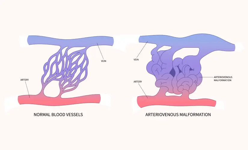
What is an Arteriovenous Malformation (AVM)?
Normally arteries connect to the veins through a network of tiny capillaries. In the AVM medical condition, blood vessels form an irregular network that creates abnormal connections between arteries and veins. AVMs lack the tiny capillaries that normally connect arteries to veins. Blood rushes straight from arteries into veins because of this abnormal structure, which bypasses a vital slowing mechanism needed to deliver oxygen to tissues and creates a high pressure zone which if ruptures can lead to Brain Hemorhage or Seizures.
Arteriovenous Malformation Symptoms
AVM symptoms often remain hidden until bleeding starts. People commonly experience:
- Seizures
- Severe headaches
- Muscle weakness or paralysis
- Loss of coordination resulting in difficulty in walking
- Numbness or tingling sensations
- Vision problems
- Dizziness
- Difficulty speaking or understanding language
- Confusion or memory issues
AVM in a newborn (called a Vein of Galen malformation) might result in:
- Enlarged head (due to fluid buildup in the brain)
- Veins of the scalp become swollen
- Seizures
Most symptoms appear between the ages of 10-40. People who reach age 50 without symptoms might never develop them.
Causes of Arteriovenous Malformation
AVMs develop during foetal growth in the womb. Scientists believe chemical imbalances that control blood vessel formation might lead to their development. Some AVMs can also develop later from injury or radiation exposure.
Risk of Arteriovenous Malformation
AVM is a rare medical condition that affects about 1 in 100,000 people. Certain factors increase AVM risk. These are:
- Men have a higher risk than women.
- People with hereditary hemorrhagic telangiectasia (HHT), also called Osler-Weber-Rendu syndrome, face greater chances of developing AVMs.
Complications of Arteriovenous Malformation
Bleeding (haemorrhage) remains the biggest risk. Serious complications include:
- Stroke
- Permanent brain damage
- Formation of aneurysms (weak bulges in blood vessels)
- Oxygen deprivation to the surrounding tissues
- Seizures
- Developmental delays
- Congestive heart failure
Diagnosis of Arteriovenous Malformation
Doctors find arteriovenous malformations through careful patient assessment. They listen for a distinctive "whooshing" sound called a bruit that blood makes as it rushes through irregular vessels.
Several imaging techniques help confirm AVMs. They are:
- DSA uses a special dye to show detailed blood vessel structures
- MRI and CT scans show subtle tissue changes and bleeding presence
- Transcranial Doppler ultrasound measures blood flow speed through vessels
Treatment for Arteriovenous Malformation
Doctors recommend three primary approaches:
- Microsurgery: This procedure removes the AVM's central portion while protecting surrounding tissues. This technique proves most effective for smaller, superficial malformations.
- Endovascular embolisation: During this procedure with the help of medical glue or coils (delivered through a catheter) your doctor blocks the AVM's blood flow.
- Stereotactic radiosurgery: This method uses focused radiation beams to target the AVM. This treatment scars and closes affected vessels gradually over one to three years like Cyber knife or Gamma Knife surgery.
When to See a Doctor
Get emergency care right away if you experience:
- Sudden, severe headache (people explain it as the worst headache in their life)
- Seizures
- Weakness in limbs
- Vision problems
- Confusion
- Memory or attention issues
Patients need quarterly checkups after treatment, followed by yearly appointments.
Conclusion
People with arteriovenous malformation face real-life challenges, yet many never show any signs of the condition. This rare condition affects just 1 in 100,000 people, which explains why public awareness remains low. However, recognising warning signs is vital since half of brain AVM cases first show up as bleeding episodes.
Today's diagnostic tools (like MRI scans and cerebral angiography) can detect these tangled blood vessels with amazing accuracy. Advanced treatment options give patients real hope to manage this rare but important medical condition well.
FAQs
1. Is an AVM serious?
Yes. Arteriovenous malformations can create serious health risks, with brain bleeding being the most important concern. Unruptured AVMs have a yearly rupture risk of 2-4%. This risk becomes much higher if the AVM has already bled once.
Many patients don't realise they have an issue until they experience a sudden, intense headache - one they often describe as the "worst headache ever." Getting immediate medical help is vital at this point.
2. At what age does AVM start?
People are born with AVMs, but they don't usually cause problems until ages 10-40. These malformations often stay quiet until adulthood. Most people discover their condition in their 40s.
Research shows that the majority of bleeding incidents happen before age 50. Some AVMs become noticeable during puberty or after someone has an accident.
3. When is surgery recommended for an arteriovenous malformation?
Doctors usually recommend immediate surgery if an AVM has already bled, because the risk of another bleed is higher. Surgery works best with smaller malformations.
The medical team looks at several factors to make their decision:
- Any previous bleeding
- The malformation's location and size
- The patient's age and overall health
- Whether there are related aneurysms
4. How often should AVMs be monitored if left untreated?
Doctors need to keep watching untreated AVMs regularly. They might suggest MRI scans or DSA every 2-5 years. This helps them spot worrying changes like new aneurysms or venous varices. Patients usually see their doctor every three months during the first year after diagnosis. After that, yearly checkups become the norm.

Still Have a Question?
