-
Doctors
-
Specialities & Treatments
Centre of Excellence
Specialties
Treatments and Procedures
Hospitals & Directions HyderabadCARE Hospitals, Banjara Hills CARE Outpatient Centre, Banjara Hills CARE Hospitals, HITEC City CARE Hospitals, Nampally Gurunanak CARE Hospitals, Musheerabad CARE Hospitals Outpatient Centre, HITEC City CARE Hospitals, Malakpet
 Raipur
 Bhubaneswar
 Visakhapatnam
 Nagpur
 Indore
 Chh. Sambhajinagar
Clinics & Medical Centers
Book an AppointmentContact Us
Online Lab Reports
Book an Appointment
Consult Super-Specialist Doctors at CARE Hospitals

Aortic Aneurysms
Aortic Aneurysms
Aortic Ulcer Treatment in Hyderabad, India
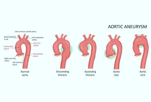
What is an Aortic Aneurysm?
It is the largest blood vessel, carrying blood from the heart to multiple parts of the body. The aorta dilates beyond 1.5 times its normal size if there is an aneurysm. An aneurysm can occur anywhere in the aorta.
The Aortic Aneurysm Treatment in Hyderabad is one of the most complex procedures performed on the human body, particularly the surgical treatment of thoracoabdominal aneurysms. A good outcome comes from a combination of factors including a host of medical professionals across a wide range of disciplines, as well as a variety of facilities. The procedure requires cardiologists, cardiovascular surgeons, interventional cardiologists, radiologists, anesthesiologists, perfusionists, physiotherapists, and dietitians as medical professionals. It is essential to have a well-equipped intensive care unit with specialist physicians as well as a blood bank capable of offering a wide variety of specialized blood products often at short notice as well as round-the-clock availability of all of these services and personnel. Due to inadequate infrastructure or lack of trained personnel, many centres avoid doing these complex operations.
At CARE Hospitals, these operations, which are the part of Aortic Aneurysm Treatment in Hyderabad, are performed in large numbers by experts who have specialized training and experience in performing this type of procedure with excellent outcomes. A multidisciplinary approach helps to tailor the most effective and safe treatment to the specific needs of each patient. CARE Hospitals has a hybrid operating room with cardiac catheterization facilities, which perform a combination of open and interventional procedures.
Types of Aortic Aneurysm
- An abdominal aortic aneurysm: It is known as an abdominal aortic aneurysm when it occurs in the abdominal part of the aorta. Aortic aneurysms treatment commonly occurs in the abdominal aorta.
- Thoracic aortic aneurysm: The thoracic aortic aneurysm occurs when the aneurysm exists in the thoracic part of the aorta.
- Thoracoabdominal aortic aneurysm: It is termed thoracoabdominal aortic aneurysm when the aneurysm occurs in both the thoracic and abdominal parts of the aorta.
- Dissecting Aortic Aneurysm: An aorta is like a sandwich with three layers. A layer known as the intima lies inside the media, which lies inside the adventitia layer.
This condition is caused when the inner layer of the aorta called intima tears and blood under high pressure separates the inner and outer layers of the media to create a false lumen causing weakness and subsequent dilatation called aortic dissection or dissecting aneurysm of the aorta.
How common are they?
Abdominal aortic aneurysms are significantly more prevalent in males and those assigned male at birth, with a ratio of 4 to 6 times compared to females and those assigned female at birth. They affect approximately 1% of men aged 55 to 64 and become increasingly common with advancing age, with the likelihood rising by around 4% every decade.
Abdominal aortic aneurysms are more common than thoracic aortic aneurysms, possibly due to the thicker and stronger wall of the thoracic aorta compared to the abdominal aorta.
Causes of aortic aneurysms
-
The process of hardening of the artery wall leads to aneurysm and dilatation.
-
An abnormality in the genetic code such as Marfan syndrome, Loeys-Dietz syndrome, and high blood pressure.
-
Infection.
-
Conditions resulting in inflammation, such as Takayasu arthritis.
-
High cholesterol
-
Trauma
-
Thoracic aortic aneurysm patients are 21 percent more likely to have a family history of this condition.
Symptoms of thoracic aortic aneurysm
-
When an aortic aneurysm is in the early stages, it is often symptomless.
-
Aneurysms of the thoracic aorta can cause chest or back pain.
-
Breathing difficulties may also occur.
-
Coughing.
-
As thoracic aortic aneurysms enlarge, the voice gets hoarse.
-
The food pipe is compressed, making swallowing difficult.
-
The aortic dissecting aneurysm causes sudden, intense back pain.
Symptoms of abdominal aortic aneurysm
The symptoms of an abdominal aortic aneurysm are abdominal pain or a pulsating abdominal lump.
The diagnosis of thoracic aortic aneurysm
-
Aneurysms of the aorta are typically asymptomatic.
-
It is most often discovered during a routine examination that you find aortic aneurysms.
-
The chest x-ray reveals large aneurysms in the thorax.
-
An echocardiogram reveals evidence of thoracic aneurysms along the ascending aortic arch and proximal descending thoracic arches.
-
Even small abdominal aortic aneurysms can be seen with an abdominal ultrasound.
-
The CT aortogram provides minute details about thoracic, thoracoabdominal, and abdominal aneurysms for treatment planning.
-
Magnetic Resonance Imaging (MRI).
-
Angiography.
Treatment of thoracic aortic aneurysms
-
Medications with control of blood pressure and 6 monthly CT or MRI follow-ups in patients with aortic aneurysms less than 5 cm in diameter with the exception of Marfan syndrome, where aortic aneurysm treatment treatment is advised regardless of aneurysm diameter.
-
TEVAR (thoracic endovascular aortic repair) is a procedure for moderately sized aortic aneurysms. Patients at high risk for open surgery can also benefit from this technique. A major advantage of this technique is the ease with which a covered metal stent can be implanted inside the aortic aneurysm through a small cut in the groin without opening the chest or abdomen. A local anaesthetic can be used to perform the procedure.
-
The surgical repair of thoracic aortic aneurysms.
Prevention
Having elevated blood pressure, high cholesterol levels, or using tobacco products heightens the likelihood of developing an aortic aneurysm. However, you can mitigate this risk by adopting a healthy lifestyle, which involves:
- Eating a heart-healthy diet.
- Getting regular exercise.
- Maintaining a healthy weight.
- Quitting smoking and using tobacco products.
Aortic Ulcer
What Is An Aortic Ulcer?
The formation of plaque by atherosclerosis causes this irregularity of the aortic wall, sometimes called a penetrating aortic ulcer. By wearing down the inner lining of the aorta, the largest blood vessel in the body that branches away from the heart, plaques cause severe damage to the blood vessel. A thoracic aortic aneurysm or aortic dissection can occur when the plaque erodes the chest artery wall.
Aortic Ulcer Symptoms
The symptoms of an aortic ulcer are difficult to describe because they are common with many other conditions. The following symptoms may indicate an aortic ulcer:
-
Aneurysm or dissection of the aorta in the family.
-
Genetic conditions that affect connective tissue, such as Marfan syndrome and Ehlers-Danlos syndrome.
-
Bicuspid aortic valve disease.
-
Coronary artery disease (heart disease), which can lead to atherosclerosis.
-
High blood pressure.
Diagnosing an Aortic Ulcer
A doctor may refer you for cardiovascular imaging tests if you complain of chest or back pain that is unusual or difficult to describe.
-
CT scan
-
MRI scan
Aortic Ulcer Treatment
The team of cardiologists, cardio surgeons and vascular surgeons at the Aortic Center lead and advance aortic disease management. The doctor wants to prevent aortic aneurysms or dissections from developing from an aortic ulcer. This is most likely accomplished by:
Activating surveillance: Often referred to as “watchful waiting,” this involves regular monitoring of the ulcer using the same imaging tests used for diagnosis.
Medications: Medications may be prescribed to treat coronary artery disease.
Depending on your doctor, you may need surgery to avoid the ulcer worsening. Surgical options may include:
-
A thoracic endovascular aortic repair (TEVAR) involves threading a metallic tube through an artery in the leg into the aorta to replace the damaged area.
-
An aorta surgery that combines endovascular (catheter-based) and open techniques to repair the wall.
-
Aorta surgery with a graft to repair the ulcerated area.
-
Aortic root replacements that spare the valve when an ulcer occurs where the aorta meets the heart are known as valve-sparing aortic root replacements.
Our Doctors
-

Dr. Tarun Gandhi
MS, FVES
Vascular & Endovascular Surgery
View More -

Dr. P C Gupta
MBBS, MS, FICA, FIVS (Japan)
Vascular & Endovascular Surgery
View More -

Dr. Ashish N Badkhal
MBBS, MS, MCh
Vascular Surgery
View More -

Dr. Ashok Reddy Somu
MBBS, MD, FVIR
Vascular & Interventional Radiology
View More -

Dr. B. Pradeep
MBBS, MD, DNB, FRCR CCT (UK)
Vascular & Interventional Radiology
View More -

Dr. Mustafa Razi
MBBS, MD
Vascular & Interventional Radiology
View More -
Dr. N. Madhavilatha
MBBS, MS, PDCC
Vascular & Endovascular Surgery
View More -

Dr. Radhika Malireddy
MBBS, DNB (General Surgery), DrNB (Plastic & Reconstructive Surgery), Post-Doctoral Fellowship in Diabetic Foot Surgery
Vascular & Endovascular Surgery
View More -

Dr. Rahul Agarwal
MBBS, DNB (General Surgery), FMAS, DrNB (Vasc. Surg)
Vascular & Endovascular Surgery
View More -

Dr. Rajesh Poosarla
MBBS, MD, DNB, DM (Gold Medalist), EBIR, FIBI, MBA (HA)
Interventional Radiology
View More -

Dr. S. Chainulu
MBBS, DNB (Radio-Diagnosis)
Vascular & Interventional Radiology
View More -

Dr. Santhosh Reddy K
MBBS, MD
Radiology
View More -

Dr. Surya Kiran Indukuri
MBBS, MS (General Surgery), DrNB (Vascular & Endovascular Surgery)
Vascular & Endovascular Surgery
View More -

Dr. Suyash Agrawal
MBBS, General Surgery (DNB), Surgical Oncology (DrNB)
Vascular & Endovascular Surgery
View More -

Dr. V. Apoorva
MBBS, MS (General Surgery), DrNB Vascular surgery
Vascular & Endovascular Surgery
View More -
Dr. Vamsi Krishna Yerramsetty
MBBS, DNB, FIVS
Vascular & Endovascular Surgery
View More -

Dr. Venugopal Kulkarni
MBBS, MS, MRCS, FRCS
Vascular & Endovascular Surgery
View More
Frequently Asked Questions
Couldn’t find what you were looking for?
Need any help? Get a Call Back.

Still Have a Question?
