-
Centre of Excellence
Specialties
Treatments and Procedures
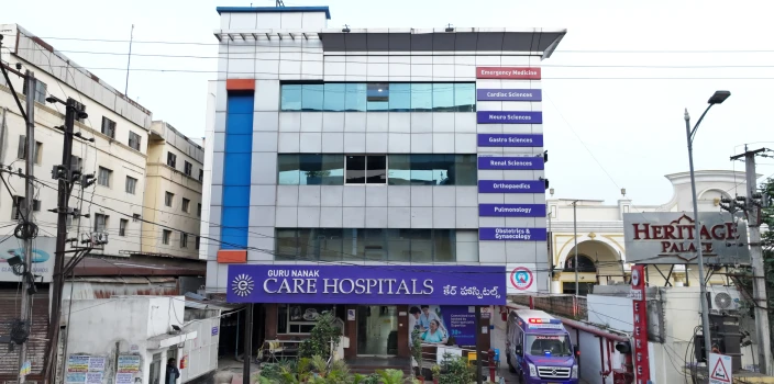
 HyderabadCARE Hospitals, Banjara Hills CARE Outpatient Centre, Banjara Hills CARE Hospitals, HITEC City CARE Hospitals, Nampally Gurunanak CARE Hospitals, Musheerabad CARE Hospitals Outpatient Centre, HITEC City CARE Hospitals, Malakpet
 Raipur
 Bhubaneswar
 Visakhapatnam
 Nagpur
 Indore
 Chh. Sambhajinagar
 Clinics & Medical Centers
-

-

-

-

-

-
Follow Us
Best Hospital for Paediatric Spinal Dysraphism Surgery in Hyderabad
- Advanced Technology
- Shorter Hospital Stay
- Pre & Post-Operative Care
- All Insurance Accepted

Chat With Our Experts
Get second opinion on Whatsapp
25 lakhs+
Happy Patients
Experienced and
skilled surgeons
17
Health Care Facilities
Top most Referral Centre
for Complex Surgeries
Advanced Paediatric Spinal Dysraphism
Paediatric spinal dysraphism is the second most common birth anomaly. The condition develops when the spine doesn't fuse correctly during embryonic development. This causes a range of structural problems that can affect the spinal cord, nerve roots, and backbone. Better nutrition for women, folic acid supplements, improved antenatal care and advanced prenatal screening have helped reduce its prevalence.
Spinal dysraphism includes both open and closed forms that need different treatment approaches. The condition affects the lumbosacral region (90% of cases), the thoracic spine, and the cervical spine. Myelomeningocele is the most common type, while meningocele and lip myelomeningocele don't happen as much. These kids often face other issues like hydrocephalus, cerebrospinal fluid leaks, and problems controlling their bowel and bladder. Early and accurate diagnosis becomes important to prevent long-term complications, especially since closed spinal dysraphism shows more subtle signs.
What is Spinal Dysraphism?
The spinal dysraphism describes the incomplete fusion or "bad suture" of the spine. The term represents many congenital malformations where the spinal cord, nerve roots, or vertebral column don't develop correctly.
Doctors see this condition very early in embryonic development. The process starts around the third week when a sheet of cells called the neural plate folds into the neural tube. The brain develops from the top portion of this tube, and the spine and spinal cord form from the rest. Spinal dysraphism results when a section of this neural tube doesn't close completely.
Best Paediatric Spinal Dysraphism Surgery Doctors in India


- No doctors found for this location.
Types of Spinal Dysraphism
Doctors group spinal dysraphism into two main categories:
- Open Spinal Dysraphism (OSD) - These defects lack skin covering and include conditions like myelomeningocele, myelocele, and posterior meningocele. Doctors call it spina bifida aperta and babies show visible abnormalities at birth.
- Closed Spinal Dysraphism (CSD) - Normal skin covers these anomalies. Signs might include a subcutaneous mass, a hairy patch, skin discolouration or a dimple. Lipomyelomeningocele, diastematomyelia, and tethered filum terminale are common examples.
Anatomical involvement varies greatly. Some forms affect only the vertebrae (spina bifida occulta). Others impact the spinal cord and its protective membranes. Split cord malformations show a unique type where the spinal cord splits lengthwise into two separate parts, sometimes separated by bone or cartilage.
The notochord plays a vital role in forming both the neural tube and various body structures. Children with spinal dysraphism often show other problems in their gastrointestinal, urogenital, or respiratory systems. This explains why VACTERL syndrome patients frequently have spinal cord malformations among other systemic issues.
Children might develop progressive neurological, urological, or orthopaedic problems as they grow.
Symptoms of Spinal Dysraphism
The spinal dysraphism symptoms vary and depend on the type and severity of the malformation. Children with this condition show various physical and neurological signs that change as they grow.
A bulging sac on the back makes open spinal dysraphism clear when a baby is born. This swelling shows up in the lower back (lumbosacral region). The surrounding skin shows discolouration or unusual texture and the bulge might have a thin, translucent membrane or exposed neural tissue.
Closed forms have more subtle external signs. These include:
- A dimple or small depression on the lower back
- Hairy patches over the spine
- Port-wine stains or unusual pigmentation
- A subcutaneous fatty mass or lump
- Skin tags or abnormal growths
Neurological symptoms:
- Children's attempts to crawl or walk reveal lower limb weakness or paralysis.
- Some children's legs move or position asymmetrically, especially when they rest
- Children's legs might show reduced sensation or abnormal reflexes
- Babies show irritability during diaper changes or when lying on their backs
- Foot deformities, including clubfoot
Urinary and bowel problems:
- Frequent urinary tract infections
- Constant wetness despite toilet training
- Unusual bowel habits
Children's growth brings more symptoms:
Older children complain about leg fatigue after minimal activity or show a decline in previously mastered motor skills.
Diagnosis of Spinal Dysraphism
A multidisciplinary assessment will give a detailed evaluation and proper management planning. Doctors use various diagnostic tools to identify and understand these complex conditions.
- Prenatal screening:
- Doctors can detect the majority of spina bifida cases and anencephaly instances through maternal serum screening.
- Ultrasound screenings at 12, 22 and 32 weeks of pregnancy help doctors spot many cases of myelomeningocele.
- Foetal MRI is a great way to get superior soft tissue visualisation that ultrasound cannot match.
- Neonatal screening: A detailed clinical examination is a vital part of the diagnosis at birth. The medical team checks the head's size, shape, fontanelles and looks for lacunar skull defects. They inspect the back's neural placode, lesion level, skin condition, and related deformities. A neurological assessment helps determine motor weakness, sensory level, and sphincter function.
- Ultrasound serves as an excellent first-line screening option for newborns with suspected occult spinal dysraphism before spinal ossification is complete.
- MRI stands as the gold standard with 100% correlation to operative findings. This method delivers exceptional soft tissue visualisation and can spot associated conditions (like tethered cord, syrinx or split cord) that ultrasound might miss.
- Doctors might also perform the following diagnostic tests:
- X-rays to check bony abnormalities
- DMSA scans so that they can know your kidneys health
- Urodynamic studies for neurogenic bladders (best done when the baby is 2-3 months old)
Treatment for Spinal Dysraphism
Each patient's treatment plan depends on the type and severity of their condition. Doctors perform surgery within 24-72 hours after birth for open spinal dysraphism. With the treatment goals including but not limited to:
- Protect neural tissue from damage
- Create a watertight dural closure
- Ensure proper skin coverage
- Prevent future tethering
Surgical techniques change based on the lesion's size & level. Surgeons first isolate the neural placode (while they preserve nerve roots), and then create a watertight dural closure.
Patients with closed spinal dysraphism might not need immediate surgery if they show no symptoms.
Nurses position patients carefully on their stomach after surgery to keep the wound clean. These children need ongoing support from physical, occupational, and recreational therapists.
Long-term care focuses on:
- Teaching bladder and bowel control through intermittent catheterisation
- Using braces to maximise mobility
- Monitoring hydrocephalus regularly
- Checking motor function
Children with spinal dysraphism can reach their full functional potential with detailed care from multiple specialists.
Conclusion
Spinal dysraphism poses a major challenge for children and families even with today's medical advances. This condition affects thousands of children yearly, though it's nowhere near as common as heart defects. The lives of these children change dramatically. Detection at an early stage makes a huge difference in outcomes, particularly for closed variants that often hide under normal-looking skin.
Parents become the first line of defence as they watch for subtle signs. Unusual skin markings, developmental delays, or changes in walking patterns might signal underlying issues. These small details could point to something significant happening under the surface.
Medical teams offer top-notch support during the entire care process. Physical therapy improves their mobility and specialised training helps with bladder and bowel management.
Best Paediatric Spinal Dysraphism Surgery Hospitals in India