-
Centre of Excellence
Specialties
Treatments and Procedures
 HyderabadCARE Hospitals, Banjara Hills CARE Outpatient Centre, Banjara Hills CARE Hospitals, HITEC City CARE Hospitals, Nampally Gurunanak CARE Hospitals, Musheerabad CARE Hospitals Outpatient Centre, HITEC City CARE Hospitals, Malakpet
 Raipur
 Bhubaneswar
 Visakhapatnam
 Nagpur
 Indore
 Chh. Sambhajinagar
 Clinics & Medical Centers
-

-

-

-

-

-
Follow Us

Best Hospital for Aneurysm Coiling in Hyderabad
- Advanced Technology
- Shorter Hospital Stay
- Pre & Post-Operative Care
- All Insurance Accepted

Chat With Our Experts
Get second opinion on Whatsapp
25 lakhs+
Happy Patients
Experienced and
skilled surgeons
17
Health Care Facilities
Top most Referral Centre
for Complex Surgeries
Advanved Aneurysm Coiling
Aneurysm coiling or endovascular coil embolisation treats intracranial aneurysms. Aneurysms are abnormal focal arterial dilations where the wall has weakened and ballooned outward. The principal danger is rupture - subarachnoid haemorrhage (SAH), which carries a higher risk of complications in untreated cases.
A microcatheter is advanced through the femoral or radial artery to the aneurysm sac. Soft platinum coils are deployed into the sac, forming a thrombogenic mesh that promotes clot formation and excludes the aneurysm from circulation eliminating rupture risk. Coiling of aneurysm is the endovascular alternative to surgical clipping.
Who Needs Aneurysm Coiling?
Coiling is indicated for both ruptured and unruptured intracranial aneurysms, selected by morphology, location, size, and surgical risk. It is recommended for:
- Ruptured aneurysms with SAH - early intervention within 24 to 72 hours prevents re-bleeding and saves patient’s life.
- Unruptured aneurysms of 4mm or larger.
- Aneurysms with high-risk morphology like irregular shape, daughter sacs, or a narrow neck
- In high surgical risk aneurysm coiling avoids craniotomy; preferred in the elderly and those with significant comorbidities.
- Posterior circulation aneurysms (basilar tip, PICA) that are anatomically difficult to clip; coiling is standard
- Patient preference for minimally invasive treatment after informed discussion.
Best Aneurysm Coiling Doctors in India


Symptoms of Brain Aneurysm
Most unruptured aneurysms are asymptomatic and found incidentally on imaging. Symptoms occur from mass effect or rupture:
- Unruptured aneurysms can cause compression of adjacent structures:
- Headache behind or above the eye; diplopia from third cranial nerve compression
- Visual field defects, facial numbness or weakness, difficulty speaking
- Ruptured aneurysms can cause a thunderclap headache:
Causes of Brain Aneurysm
Brain aneurysms usually develop when a weak area in the wall of a blood vessel (predominantly bifurcations in the circle of Willis) gradually bulges outward over time. Several factors can contribute to this weakening of the artery wall. These are:
- Congenital vessel wall (tunica media) weakness at bifurcation points
- Long-term uncontrolled hypertension accelerates wall stress and degeneration
- Atherosclerosis in blood vessels can weaken the artery walls over time
- Mycotic aneurysms from infective endocarditis can occasionally lead to infected aneurysms
- Flow-related aneurysms on arteriovenous malformations (AVMs) can alter blood flow and increase stress on nearby arteries.
Risk Factors for Brain Aneurysm
Several lifestyle, medical, and genetic factors can increase the likelihood of developing a brain aneurysm or aneurysm rupture:
- Hypertension: The leading modifiable risk factor and poorly controlled blood pressure accelerates growth and rupture risk
- Smoking: Tobacco use significantly increases the chances of aneurysm formation, growth and rupture
- Family history: First-degree relatives carry three to seven times population risk
- Female sex: Brain aneurysms are more commonly seen in women, especially with increasing age
- Connective tissue disorders: ADPKD, Ehlers-Danlos syndrome type IV, and Marfan syndrome
- Prior aneurysm: Individuals who have had one aneurysm may be at risk of developing another
- Substance abuse: Cocaine and certain stimulant drugs can sharply increase blood pressure and trigger aneurysm rupture.
Complications of Aneurysm Coiling
Aneurysm coiling is safe at experienced neurovascular centers and when performed by qualified Neurointervention specialist ( Interventional Neuroradiologist) but complications can arise. They are:
- Peri-procedural thromboembolic stroke
- Aneurysm perforation
- Coil compaction and recanalisation
- Vasospasm in SAH
- Access site complications
- Contrast nephropathy.
Diagnosis of Brain Aneurysm
The diagnostic pathway differs for ruptured and unruptured presentations:
- Neurological Examination: Doctors assess symptoms such as headache, vision changes, weakness, speech difficulty, or altered consciousness
- Non-contrast CT brain: Detects subarachnoid blood in the majority of cases.
- Lumbar puncture: If CT is negative, spinal fluid testing confirms SAH from 12 hours to 2 weeks post-rupture
- CT Angiography (CTA): This rapid, non-invasive test detects aneurysms of 3 mm or larger (performed immediately in suspected SAH)
- MR Angiography (MRA): Preferred for screening at-risk patients and post-coiling surveillance
- Digital Subtraction Angiography (DSA): Definitive imaging and is performed immediately before coiling for the highest-resolution anatomy.
Aneurysm Coiling Procedure
Performed under general anaesthesia with systemic heparinisation. Procedural steps:
- Doctors make a femoral access and guide a catheter toward the internal carotid or vertebral artery
- Interventional Neuroradiologist performs a diagnostic angiography to confirm size, neck width, dome-to-neck ratio, and parent artery relationship that guides coil selection
- A very thin microcatheter is carefully advanced into the aneurysm under imaging guidance
- Coils are deployed in the aneurysm to fill the weakened area and block abnormal blood flow
- In some cases, like wide-necked aneurysms require temporary balloon remodelling or parent artery stenting to prevent coil herniation
- Completion angiography is performed to confirm successful sealing of the aneurysm and parent artery patency
The procedure usually takes one to three hours. Post-procedure monitoring in the neurosurgical ICU with vigilance for neurological deterioration, vasospasm and access site bleeding.
Benefits of Aneurysm Coiling
Aneurysm coiling offers a minimally invasive way to treat brain aneurysms:
- Minimally invasive procedure without open brain surgery
- Lower surgical site infection, brain injury and anesthetic complications
- Superior outcomes in ruptured aneurysms in emergency.
- Faster recovery - unruptured aneurysm patients discharged within 24 to 48 hours
- Quicker functional return than, after craniotomy
- High surgical risk patients tolerate endovascular treatment better than open surgery
- Posterior circulation and deep aneurysms technically easier by the endovascular route
- Immediate re-bleeding prevention in ruptured SAH
- Improves long-term neurological safety and quality of life.
When to See a Doctor
Consult a doctor immediately if:
- Headache described as the worst of one's life
- Sudden double vision or drooping eyelid
- Sudden loss of consciousness or new-onset seizure
Known unruptured aneurysm patients should seek review if headache character changes or neurological symptoms develop. High-risk individuals (ADPKD, strong family history) should discuss screening MRA with their specialist.
Conclusion
Aneurysm coiling has transformed intracranial aneurysm treatment as effective, durable occlusion without craniotomy. In ruptured SAH, the studies demonstrate superior one-year outcomes versus surgical clipping for anatomically suitable aneurysms. Case selection by an experienced neurovascular MDT remains essential.
In a ruptured aneurysm, re-bleeding is the most preventable cause of complications and coiling within 24 hours reduces that risk. In unruptured aneurysm, the treat-versus-surveil decision is individual. Both benefit from early specialist input rather than deferred referral.
Aneurysm Coiling Hospitals in India
-
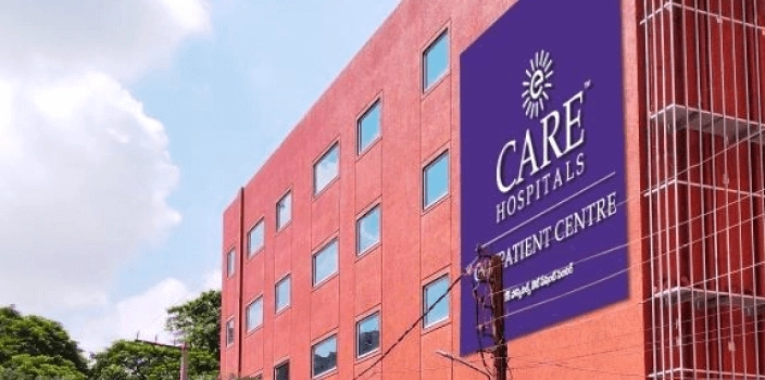
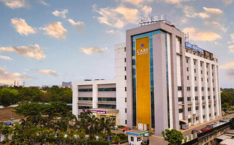
CARE Hospitals, Banjara Hills, Hyderabad
CARE Hospitals Outpatient Centre, Banjara Hills, Hyderabad
CARE Hospitals, HITEC City, Hyderabad
CARE Hospitals Outpatient Centre, HITEC City, Hyderabad
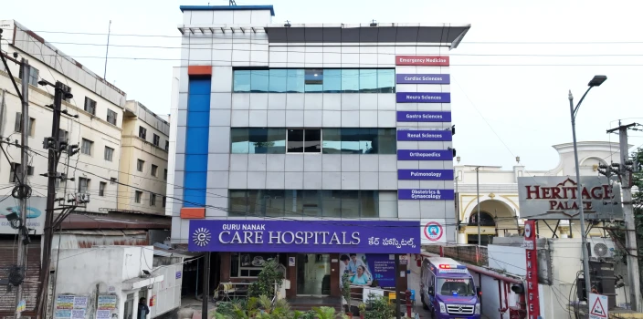
Gurunanak CARE Hospitals, Musheerabad, Hyderabad
CARE Hospitals, Nampally, Hyderabad
CARE Hospitals, Malakpet, Hyderabad
CARE Hospitals, Bhubaneswar
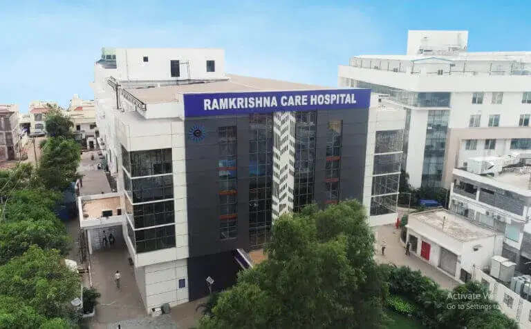
Ramkrishna CARE Hospitals, Raipur
CARE Hospitals, Ramnagar, Visakhapatnam
CARE Hospitals, Health City, Arilova
Frequently Asked Questions
Coiling treats intracranial aneurysms to prevent rupture or stop re-bleeding after rupture. It is used for both ruptured aneurysms (emergency treatment to prevent fatal re-haemorrhage) and unruptured aneurysms in high-risk patients where estimated rupture risk justifies intervention over surveillance.
Aneurysm coiling and open brain surgery (surgical clipping) are both effective treatments for ruptured aneurysms. Coiling is minimally invasive and is often preferred because it usually involves faster recovery, shorter hospital stay, and less surgical trauma. For unruptured aneurysms, the choice depends on morphology like wide-necked aneurysms are often better clipped; posterior circulation aneurysms are almost always coiled.
Typically 1 to 3 hours depending on aneurysm complexity, neck width, and whether balloon or stent assistance is required. Emergency cases in ruptured SAH are performed as soon as possible.
It is a major interventional procedure with general anaesthesia, ICU monitoring, and recognised serious complication rates. But there is no craniotomy, skull opening, or brain retraction. The entire procedure is performed through arterial catheterisation from the groin.
For elective (unruptured) coiling, discharge is within 24 to 48 hours and you can resume light activities within one to two weeks. For ruptured SAH, ICU stay is typically 10 to 21 days, with rehabilitation in required patients. Recovery depends on SAH grade at presentation and vasospasm severity.
Common risks are:
- Peri-procedural stroke
- Accidental aneurysm perforation
- Coil compaction with recanalisation
In SAH, vasospasm-related delayed ischaemia (days 4 to 14) is the major post-procedure risk. Structured follow-up imaging is required for all patients.
Any cerebral aneurysm can be treated by endovascular approach. Favorable features include narrow-necked aneurysms, posterior circulation location, any age, and high surgical risk. Wide-necked aneurysms can be coiled with balloon or stent assistance. Very small asymptomatic aneurysms in low-risk patients may be surveilled rather than treated. A neurovascular multidisciplinary team assessment determines the approach.
Complete occlusion is achieved in 90% at the time of coiling. Coil compaction and recanalization occur particularly in aneurysms larger than 10 mm or with wide necks. Surveillance MRA or DSA is performed at 6 months, 18 months, and 5 years.
Still Have a Question?