-
Centre of Excellence
Specialties
Treatments and Procedures
 HyderabadCARE Hospitals, Banjara Hills CARE Outpatient Centre, Banjara Hills CARE Hospitals, HITEC City CARE Hospitals, Nampally Gurunanak CARE Hospitals, Musheerabad CARE Hospitals Outpatient Centre, HITEC City CARE Hospitals, Malakpet
 Raipur
 Bhubaneswar
 Visakhapatnam
 Nagpur
 Indore
 Chh. Sambhajinagar
 Clinics & Medical Centers
-

-

-

-

-

-
Follow Us

Best Hospital for Fracture Treatment in Bhubaneswar
- Advanced Technology
- Shorter Hospital Stay
- Pre & Post-Operative Care
- All Insurance Accepted

Chat With Our Experts
Get second opinion on Whatsapp
25 lakhs+
Happy Patients
Experienced and
skilled surgeons
17
Health Care Facilities
Top most Referral Centre
for Complex Surgeries
Advanced Fracture Treatment in Bhubaneswar
Fracture management spans a broader clinical range than any single orthopaedic subspecialty ranging from closed manipulation under anaesthesia for a displaced distal radius fracture to multi-stage reconstruction for an open tibial diaphyseal injury with bone loss. Treatment selection is tailored to the fracture pattern, soft-tissue status, bone quality, patient age, and the anatomical demands of the injured segment. CARE Hospitals, Bhubaneswar, provides the complete spectrum of fracture care, including emergency stabilisation, definitive fixation, and functional rehabilitation within a single orthopaedic unit.
What is Fracture Treatment?
Fracture treatment restores bone continuity, corrects deformity and protects the injured limb until union is achieved. Your doctor does it through operative or non-operative means (depending on fracture stability, displacement, and functional requirements). Non-operative management with cast immobilisation remains appropriate for undisplaced or minimally displaced fractures with acceptable alignment. However displaced, unstable, intra-articular, or open fractures require surgical reduction and internal or external fixation to restore anatomy, prevent malunion, and permit early joint mobilisation.
Best Fracture Treatment Doctor in Bhubaneswar

Types of Fractures Treated
Fractures are of the following types:
- Diaphyseal fractures: Involves the shaft (middle portion) of long bones—your femur, tibia and humerus
- Periarticular fractures: Occur near a joint like distal femur, tibial plateau, distal tibia (pilon), and calcaneus
- Intra-articular fractures: Extend into the joint space like the tibial plateau (Schatzker I–VI), distal humerus, radial head, and acetabulum
- Distal radius fractures: Involves the wrist end of the radius
- Vertebral fractures: Involves the vertebrae (bones of the spine)
- Stress fractures: Repetitive stress can cause small cracks (tibial anterior cortex, femoral neck, and fifth metatarsal base are at high non-union risk)
- Close fractures: Bone breaks without piercing the skin.
- Open fractures: Bone fragments can penetrate the skin and have a higher risk of infection.
- Greenstick Fracture: It is a partial bone break and is common in children.
- Pathological fractures: Occur in weakened bones due to diseases like metastatic bone disease, primary bone tumour, or osteoporotic insufficiency
Causes of Fractures
Fracture mechanism determines displacement pattern, associated soft tissue injury, and fixation strategy. Principal mechanisms are:
- High-energy trauma: Road traffic accidents, industrial injuries, and falls from height producing comminuted diaphyseal and periarticular fractures with significant soft tissue stripping
- Low-energy falls: The predominant mechanism in osteoporotic fractures of the distal radius, proximal humerus, vertebral body, and proximal femur in patients over 65
- Sports-related forces: Avulsion fractures at tendon insertion sites, stress fractures from repetitive loading and acute periarticular fractures from contact and torsional injury
- Pathological bone: Fractures through osteolytic metastases, primary bone tumours, or Paget's disease occurring with minimal or no trauma
Diagnosis of Fractures
Clinical assessment identifies deformity, neurovascular deficit and open wound, and imaging characterises fracture geometry and forms the main pillars based on which your doctor plans fixation:
- X-ray: Anteroposterior and lateral views in two planes at minimum (with traction view for comminuted articular fractures, your doctor can assess fragment displacement)
- CT scan: Detects intra-articular fractures and is helpful in three-dimensional reconstruction for surgical templating and screw trajectory planning
- MRI: Identifies occult fractures not visible on plain radiographs (undisplaced femoral neck stress fractures & bone marrow contusion patterns in knee injuries)
- Neurovascular assessment: Your doctor can recommend an ankle-brachial pressure index (ABPI) for tibial and femoral fractures with suspected vascular injury. Nerve conduction studies detect associated peripheral nerve injury
- Bone density scan (DEXA): Indicated for low-energy fragility fractures.
Fracture Treatment Procedure
Emergency management:
- Open fractures receive emergency irrigation with three litres of normal saline and surgical debridement within 6 hours of injury, as the window beyond which infection risk rises sharply. Neurovascular compromise from fracture displacement is treated by urgent reduction, and vascular injury confirmed on CT angiography requires vascular surgical intervention before orthopaedic fixation. Spanning external fixation stabilises comminuted periarticular fractures until soft tissue swelling permits definitive internal fixation (generally for 5 to 10 days).
Surgery:
- Intramedullary nailing: The fixation standard for diaphyseal fractures of the femur, tibia and humerus. The doctor inserts a locked titanium nail into the intramedullary canal through a small proximal or distal entry portal under fluoroscopic guidance, and interlocking screws proximally and distally control rotation and length. Allows immediate weight bearing in femoral and tibial nailing where comminution permits.
- Locking plate fixation: Periarticular and metaphyseal fractures (distal femur, proximal tibia, distal radius, proximal humerus, calcaneus) are fixed with anatomically precontoured locking plates. Locking screw plate construct functions as a fixed-angle device critical in osteoporotic bone (where conventional screw purchase is inadequate).
- Percutaneous screw fixation: Minimally displaced intra-articular fractures fixed with cannulated screws inserted over guidewires under fluoroscopic control.
- External fixation: Temporary spanning fixation for open fractures and periarticular injuries with severe soft tissue compromise.
- Non-operative management: Undisplaced clavicle fractures, stable vertebral compression fractures, and minimally displaced distal radius fractures in low-demand elderly patients managed with cast immobilisation, functional bracing or sling support where alignment is acceptable.
Recovery after Fracture Treatment
Rehabilitation timelines are fracture- and fixation-specific. Femoral and tibial intramedullary nail patients begin partial weight-bearing within 24 to 48 hours. Distal radius volar plate patients commence active wrist motion at two weeks. Tibial plateau fixation restricts weight-bearing for six to twelve weeks depending on fracture severity and fixation construct stability. Physiotherapy like joint mobilisation, periarticular strengthening, and proprioceptive re-education is initiated within the first week across all fixation types where soft tissue conditions permit.
Why Choose CARE Hospitals for Fracture Treatment
CARE Hospitals, Bhubaneswar, manages fractures across the full severity spectrum ranging from isolated closed injuries to complex open fractures and polytrauma within a single orthopaedic trauma unit. The department operates with 24-hour emergency orthopaedic cover, a dedicated trauma theatre with image intensifier fluoroscopy and a full implant inventory, intramedullary nails, locking plates, cannulated screws and circular frame systems. Surgeons hold postgraduate orthopaedic training with subspecialty experience in trauma reconstruction.
Complex periarticular fractures like tibial plateau, acetabulum, pilon, and calcaneus are managed with pre-operative CT three dimensional planning and intra operative fluoroscopic verification of reduction and implant position. Post-operative follow-up at two weeks, six weeks, three months and six months includes radiographic assessment of union and functional outcome scoring to identify complications at the earliest stage amenable to intervention.
Conclusion
No two fractures are quite the same (and that's exactly why treatment can't be one size fits all). The right approach depends on where the fracture is, how severe it is and the overall health of the patient. Some fractures respond well to immobilisation and a structured rehabilitation programme. Others (particularly complex or displaced ones) need surgical intervention to properly realign the bone and restore stability before healing can begin.
The reassuring part is that with the right care and a personalised treatment plan, most patients do regain their mobility and get back to living normally. At CARE Hospitals, fracture care is built on two pillars working together: advanced surgical expertise and integrated rehabilitation. Because treating the fracture is only part of the journey. Helping you recover fully, safely, and confidently is the other.
Fracture Treatment Hospitals in India
-
CARE Hospitals, Banjara Hills, Hyderabad
CARE Hospitals Outpatient Centre, Banjara Hills, Hyderabad
CARE Hospitals, HITEC City, Hyderabad
CARE Hospitals Outpatient Centre, HITEC City, Hyderabad
Gurunanak CARE Hospitals, Musheerabad, Hyderabad
CARE Hospitals, Nampally, Hyderabad
CARE Hospitals, Malakpet, Hyderabad
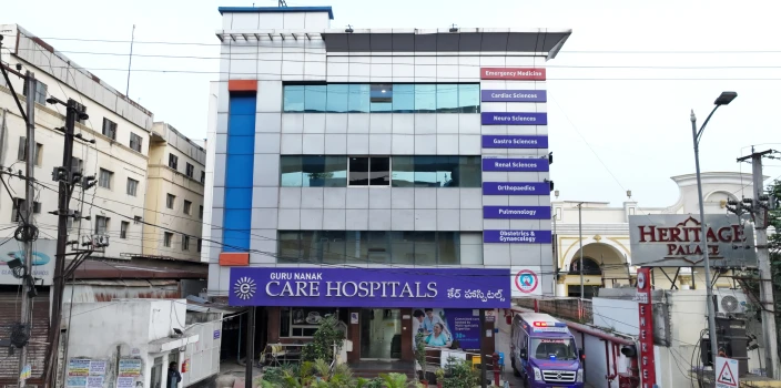
CARE Hospitals, Bhubaneswar
Ramkrishna CARE Hospitals, Raipur
CARE Hospitals, Ramnagar, Visakhapatnam
CARE Hospitals, Health City, Arilova
Frequently Asked Questions
Union time varies by fracture site, patient age, bone quality and fixation method. Simple fractures consolidate radiographically at six to eight weeks. However diaphyseal fractures require twelve to twenty weeks.
No. Undisplaced fractures, stable compression fractures of the vertebral body, and fractures in anatomically acceptable alignment in low demand patients are managed nonoperatively. Surgical fixation is reserved for displaced or unstable fractures, intra-articular injuries where articular step exceeds 2 mm, open fractures requiring debridement and fractures associated with neurovascular compromise requiring urgent reduction.
An open fracture is one where the bone communicates with the external environment through a skin wound - either from the bone end penetrating outward or a traumatic laceration exposing the fracture site. Bacterial contamination begins at the time of injury. Therefore surgical debridement within six hours reduces deep infection rates. Delay beyond this window substantially increases the risk of osteomyelitis and non-union.
Delayed union is defined as failure to achieve radiographic bridging callus by the expected timeframe for the fracture site - typically sixteen weeks for tibial diaphyseal fractures. It is distinct from non-union, where healing has ceased entirely. Management options include dynamisation of an intramedullary nail to increase compressive loading at the fracture site, exchange nailing with a larger diameter implant, bone grafting, or adjunctive low-intensity pulsed ultrasound (LIPUS) therapy.
Still Have a Question?